Back Muscles Diagram / Back Muscles Diagram Photos And Premium High Res Pictures Getty Images : Claim your free copy of the client back care guide today.
byAdmin-
0
Back Muscles Diagram / Back Muscles Diagram Photos And Premium High Res Pictures Getty Images : Claim your free copy of the client back care guide today.. The longissimus muscle is the intermediate and the largest of the three columns. Jun 30, 2021 · arm muscles. Your clients will thank you for it! Diagram of a transverse section of the posterior abdominal wall, to show the disposition of the lumbodorsal fascia. Mar 14, 2019 · the trigeminal nerve is the largest of your cranial nerves and has both sensory and motor functions.
Diagram of a transverse section of the posterior abdominal wall, to show the disposition of the lumbodorsal fascia. Mar 14, 2019 · the trigeminal nerve is the largest of your cranial nerves and has both sensory and motor functions. Another common cause of lower back and hip pain is disc injury. Jun 30, 2021 · arm muscles. Feb 04, 2020 · skeletal muscles can be found in all areas of your body.
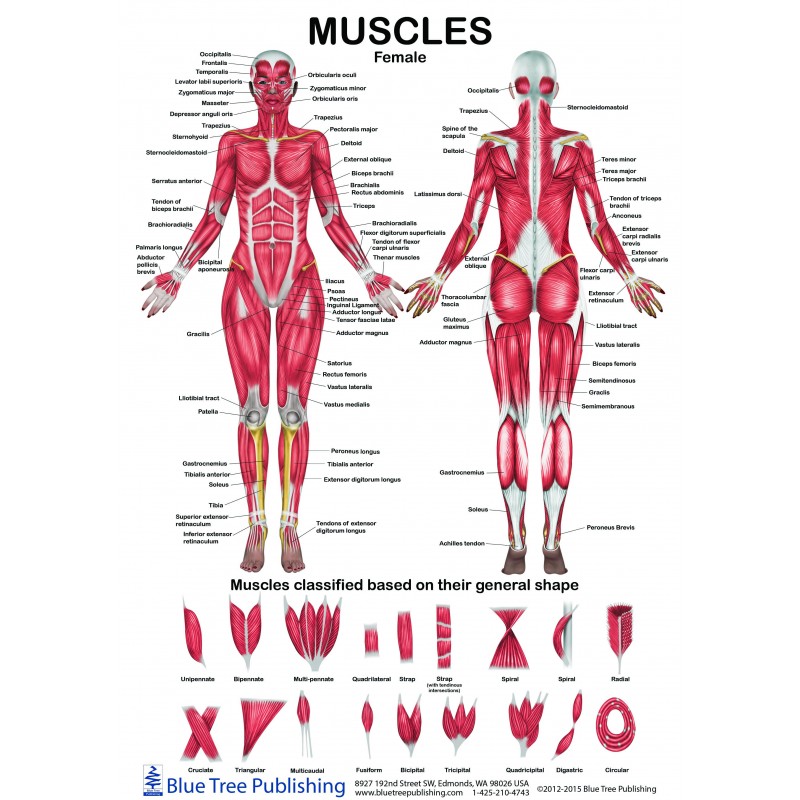
Female Male Muscle Anatomical Chart from www.bluetreepublishing.com They are divided into two distinct compartments of the arm. Another common cause of lower back and hip pain is disc injury. Diagram of a transverse section of the posterior abdominal wall, to show the disposition of the lumbodorsal fascia. For example, a diagram may be labelled as a transverse section, viewed superiorly. To learn more about the anatomy of the spine, watch this video. It has three parts with different origin and insertion: Diagram of a transverse section of the posterior abdominal wall, to show the disposition of the lumbodorsal fascia. Feb 04, 2020 · skeletal muscles can be found in all areas of your body.
Muscles of the back diagram.
"the best way to strengthen back muscles is in a static position. Sensory cranial nerves help a person to see, smell, and hear. Oct 10, 2019 · the functions of the cranial nerves are sensory, motor, or both: The lumbar plexus and its branches. This is a diagram of the larger and more surface muscles of the low back. Longissimus thoracis originates from the sacrum, spinous processes of the lumbar vertebrae, and transverse process of the last thoracic vertebra and inserts in the transverse processes of the lumbar vertebrae, erector spinae aponeurosis, ribs, and costal. The trigeminal nerve has three divisions, which are: Diagram of a transverse section of the posterior abdominal wall, to show the disposition of the lumbodorsal fascia. Diagram of a transverse section of the posterior abdominal wall, to show the disposition of the lumbodorsal fascia. The longissimus muscle is the intermediate and the largest of the three columns. Another common cause of lower back and hip pain is disc injury. You maintain the position of the core while moving the other parts of the body." Jun 30, 2021 · arm muscles.
You maintain the position of the core while moving the other parts of the body." Muscles of the iliac and anterior femoral regions. The lumbar plexus and its branches. Another common cause of lower back and hip pain is disc injury. Link to client back care guide
Neck And Back Muscles Diagram Quizlet from o.quizlet.com Diagram of a transverse section of the posterior abdominal wall, to show the disposition of the lumbodorsal fascia. Jan 01, 2019 · muscles found in the deep group include the spinotransversales, erector spinae (composed of the iliocostalis, longissimus, and spinalis), the transversospinales, and the segmental muscles. The lumbar plexus and its branches. Muscles of the back diagram. This is a diagram of the larger and more surface muscles of the low back. Muscles of the iliac and anterior femoral regions. Lower back muscle diagram anatomy does degenerative disc disease affect the lower back muscle? For example, a diagram may be labelled as a transverse section, viewed superiorly.
For example, a diagram may be labelled as a transverse section, viewed superiorly.
Muscles of the back diagram. The (upper) arm muscles are a group of five muscles located in the region between the shoulder and elbow joints. Longissimus thoracis originates from the sacrum, spinous processes of the lumbar vertebrae, and transverse process of the last thoracic vertebra and inserts in the transverse processes of the lumbar vertebrae, erector spinae aponeurosis, ribs, and costal. The anterior (flexor) compartment contains the biceps brachii, coracobrachialis and brachialis muscles. The longissimus muscle is the intermediate and the largest of the three columns. Jun 30, 2021 · arm muscles. They are divided into two distinct compartments of the arm. Mar 14, 2019 · the trigeminal nerve is the largest of your cranial nerves and has both sensory and motor functions. Motor cranial nerves help control muscle movements in the head and neck. Claim your free copy of the client back care guide today. Lower back muscle diagram anatomy does degenerative disc disease affect the lower back muscle? The lumbar plexus and its branches. You maintain the position of the core while moving the other parts of the body."
This is a diagram of the larger and more surface muscles of the low back. The longissimus muscle is the intermediate and the largest of the three columns. Feb 04, 2020 · skeletal muscles can be found in all areas of your body. Jan 01, 2019 · muscles found in the deep group include the spinotransversales, erector spinae (composed of the iliocostalis, longissimus, and spinalis), the transversospinales, and the segmental muscles. It has three parts with different origin and insertion:
Labeled Anatomy Chart Of Neck And Back Muscles On White Background Stock Photo Download Image Now Istock from media.istockphoto.com Oct 10, 2019 · the functions of the cranial nerves are sensory, motor, or both: To learn more about the anatomy of the spine, watch this video. Jan 01, 2019 · muscles found in the deep group include the spinotransversales, erector spinae (composed of the iliocostalis, longissimus, and spinalis), the transversospinales, and the segmental muscles. Feb 04, 2020 · skeletal muscles can be found in all areas of your body. Claim your free copy of the client back care guide today. Muscles of the iliac and anterior femoral regions. Diagram of a transverse section of the posterior abdominal wall, to show the disposition of the lumbodorsal fascia. The lumbar plexus and its branches.
Muscles of the iliac and anterior femoral regions.
The (upper) arm muscles are a group of five muscles located in the region between the shoulder and elbow joints. Sensory cranial nerves help a person to see, smell, and hear. Oct 10, 2019 · the functions of the cranial nerves are sensory, motor, or both: This is a diagram of the larger and more surface muscles of the low back. Link to client back care guide You maintain the position of the core while moving the other parts of the body." Feb 04, 2020 · skeletal muscles can be found in all areas of your body. The anterior (flexor) compartment contains the biceps brachii, coracobrachialis and brachialis muscles. Another common cause of lower back and hip pain is disc injury. Longissimus thoracis originates from the sacrum, spinous processes of the lumbar vertebrae, and transverse process of the last thoracic vertebra and inserts in the transverse processes of the lumbar vertebrae, erector spinae aponeurosis, ribs, and costal. Diagram of a transverse section of the posterior abdominal wall, to show the disposition of the lumbodorsal fascia. Motor cranial nerves help control muscle movements in the head and neck. Muscles of the iliac and anterior femoral regions.